Fetal Kidney Size Chart

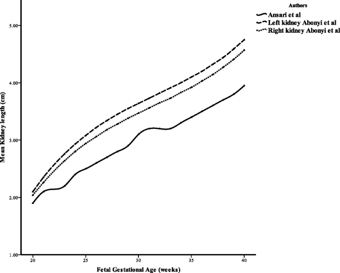
Ultrasound Measurement Of Fetal Kidney Length In Normal Pregnancy

Ultrasound Measurement Of Fetal Kidney Length In Normal Pregnancy

Table 1 From Fetal Kidney Measurement In 26 39 Weeks Gestation In

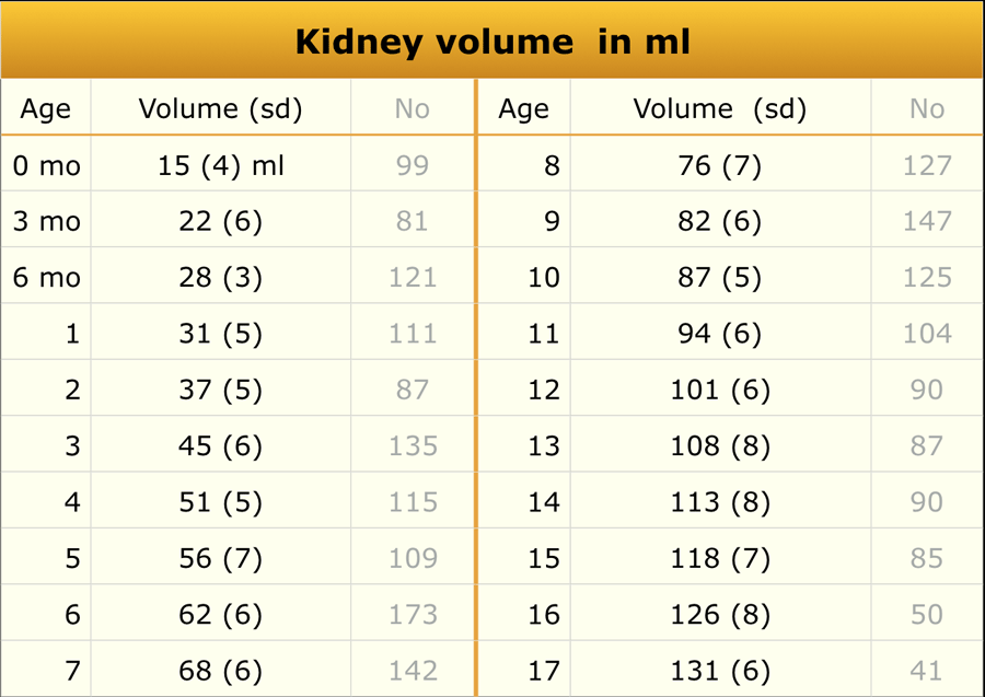
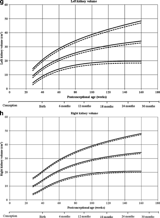
Size And Volume Charts Of Fetal Kidney Renal Pelvis And Adrenal

Reference Ranges For Ultrasound Measurements Of Fetal Kidneys In A

Ultrasound Measurement Of Fetal Kidney Length In Normal Pregnancy

I Relationship Between Volume Of Left Fetal Kidney S Cortex And

Measurement Of Renal Dimensions In Vivo A Critical Appraisal

Table 4 From Fetal Kidney Measurement In 26 39 Weeks Gestation In

Table 2 From Fetal Kidney Measurement In 26 39 Weeks Gestation In

Measurement Of Renal Dimensions In Vivo A Critical Appraisal
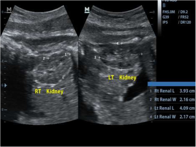
The Radiology Assistant Normal Values Ultrasound

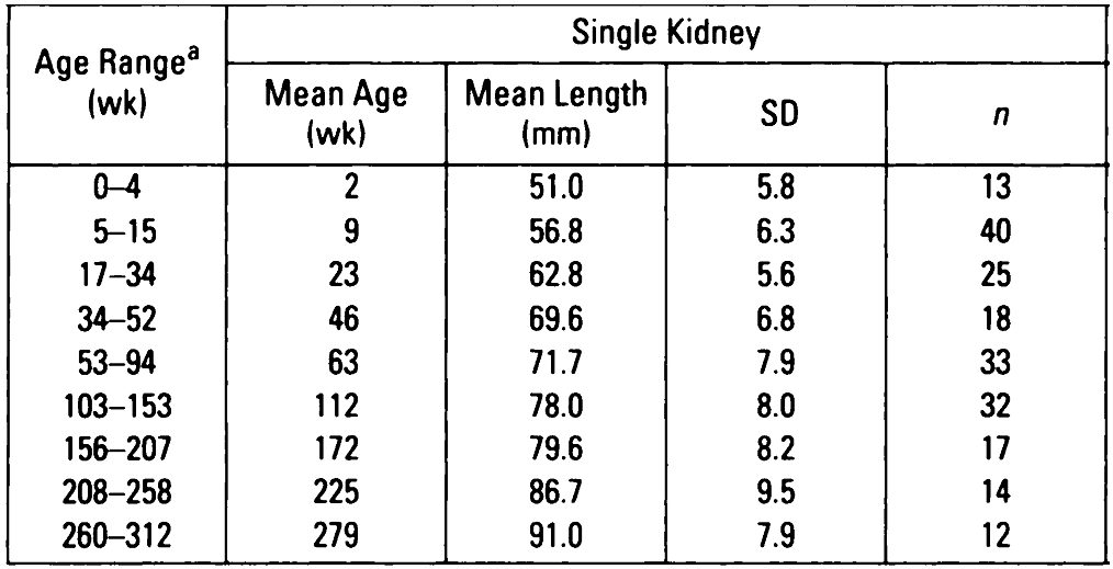
Compensatory Enlargement Of A Solitary Functioning Kidney During
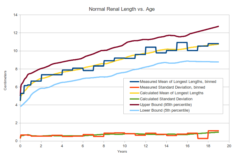
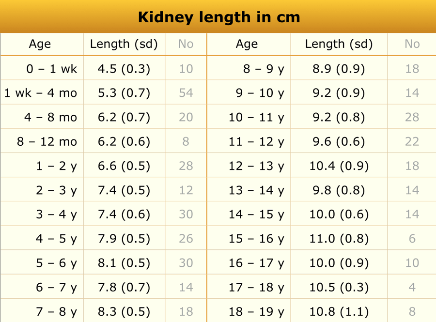
Pediatric Kidney Size Normal Range And Renal Length Percentile
Fetal Kidney Measurement In 26 39 Weeks Gestation In Normal

Sonographic Assessment Of Renal Growth In Patients With Beckwith

Size And Volume Charts Of Fetal Kidney Renal Pelvis And Adrenal

Age Related Changes Of The Human Fetal Kidney Size Semantic Scholar
Renal Anomalies Detected Or Suspected Antenatally
Https Www Cfef Org Boite A Outils Images Hauteursurrenale2014 Pdf

I Relationship Between Volume Of Left Fetal Kidney S Cortex And

Growth Curves Of Fetal Kidneys From 21 To 27 Weeks Of Gestation
Gale Onefile Health And Medicine Document Assessment Of
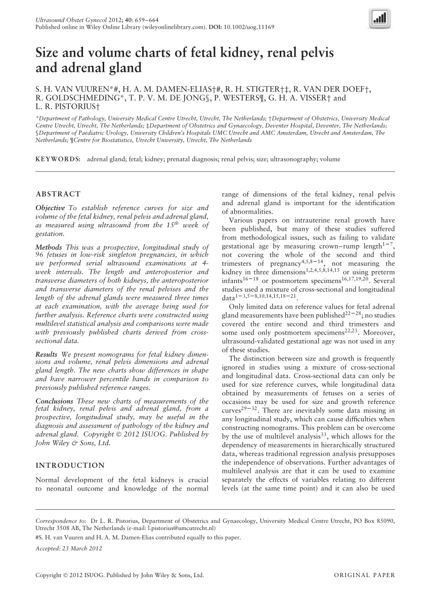
Pdf Size And Volume Charts Of Fetal Kidney Renal Pelvis And
Https Encrypted Tbn0 Gstatic Com Images Q Tbn 3aand9gcqnqf4cjegdx4m6dvf5ndemzaw Itto7xkrsl5eiu4naaofkjom Usqp Cau
Sonographic Estimation Of Gestational Age From 20 To 40 Weeks By
Https Obgyn Onlinelibrary Wiley Com Doi Pdf 10 1002 Pd 693

Normal Ultrasound Dimensions Of Newborn Kidneys In Southwest

Predicted Values Of Foot Length By Week Of Fetal Age From Linear
Sonographic Estimation Of Gestational Age From 20 To 40 Weeks By

Measurement Of Renal Dimensions In Vivo A Critical Appraisal
Mild To Moderate Renal Pelvis Dilatation Identified During
Https Onlinelibrary Wiley Com Doi Pdf 10 7863 Jum 1988 7 4 197
The Radiology Assistant Normal Values Ultrasound
Fetal Kidney Measurement In 26 39 Weeks Gestation In Normal

Mean Fetal Weight Kidney Volume And Kv Fw Ratio In Total Sample
Https Obgyn Onlinelibrary Wiley Com Doi Pdf 10 1002 Pd 693

Ultrasound Appearance Of Normal Fetal Kidneys At 32 Weeks

Pdf Prenatal Natural History Of Isolated Fetal Mild Bilateral
Sonographic Estimation Of Gestational Age From 20 To 40 Weeks By
Kidney Growth Curves In Healthy Children From The Third Trimester

Study Of Fetal Kidney Size By Ultrasonography At Different

Sonographic Assessment Of Renal Growth In Patients With Beckwith

Fetal And Infant Growth Patterns And Kidney Function At School Age
Sonographic Estimation Of Gestational Age From 20 To 40 Weeks By

Study Of Fetal Kidney Size By Ultrasonography At Different
Https Www Mdpi Com 2077 0383 7 10 324 Pdf

Normal Ultrasound Dimensions Of Newborn Kidneys In Southwest

Ultrasound Measurement Of Fetal Kidney Length In Normal Pregnancy
Https Encrypted Tbn0 Gstatic Com Images Q Tbn 3aand9gcq8u Ijlnytxehtejigehq 0taikuivy6hseyp9o6qcwgspespo Usqp Cau
Mild To Moderate Renal Pelvis Dilatation Identified During
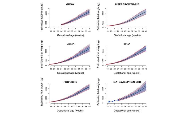
Which Fetal Size Standard Should Be Used For Diagnosing A Small

Evaluation Of Fetal Kidney Growth Using Ultrasound A Systematic

Fetal And Infant Growth Patterns And Kidney Function At School Age

Comparative Effectiveness Of A Pilot Patient Centered Ultrasound

Study Of Fetal Kidney Size By Ultrasonography At Different
Http Iaimjournal Com Wp Content Uploads 2016 08 Iaim 2016 0308 06 Pdf
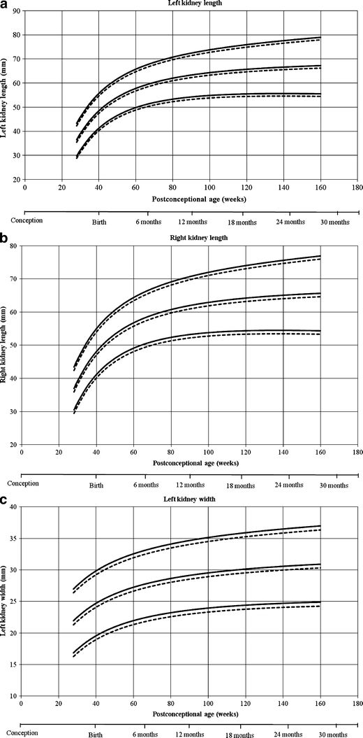
Kidney Growth Curves In Healthy Children From The Third Trimester
Fetal Kidney Measurement In 26 39 Weeks Gestation In Normal

Outcome Of Fetal Renal Pelvic Dilatation Diagnosed During The
Mild To Moderate Renal Pelvis Dilatation Identified During
Sonographic Estimation Of Gestational Age From 20 To 40 Weeks By

Measurement Of Renal Dimensions In Vivo A Critical Appraisal
Fetal Kidney Measurement In 26 39 Weeks Gestation In Normal

Nomogram Of Fetal Renal Growth Expressed In Length And Parenchymal

Normal 1st Trimester Ultrasound How To

Fetal Genitourinary Tract Radiology Key

Revised Guidelines On Management Of Antenatal Hydronephrosis Sinha

Study Of Fetal Kidney Size By Ultrasonography At Different
Mild To Moderate Renal Pelvis Dilatation Identified During

Mild Fetal Renal Pelvis Dilatation Much Ado About Nothing

Kidney Growth Curves In Healthy Children From The Third Trimester
Https Onlinelibrary Wiley Com Doi Pdf 10 7863 Jum 1982 1 7 265
Fetal Development Embryology
Https Encrypted Tbn0 Gstatic Com Images Q Tbn 3aand9gcq8u Ijlnytxehtejigehq 0taikuivy6hseyp9o6qcwgspespo Usqp Cau

Study Of Fetal Kidney Size By Ultrasonography At Different

Dissecting The Global Dynamic Molecular Profiles Of Human Fetal

Normal 1st Trimester Ultrasound How To

Sonographic Assessment Of Renal Growth In Patients With Beckwith
The Radiology Assistant Normal Values Ultrasound
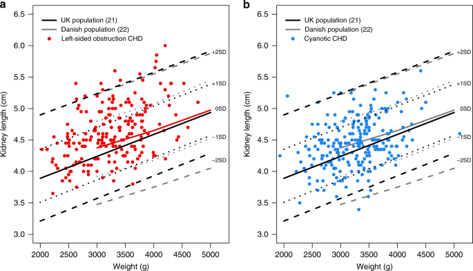
Altered In Utero Kidney Development In Newborns With Congenital
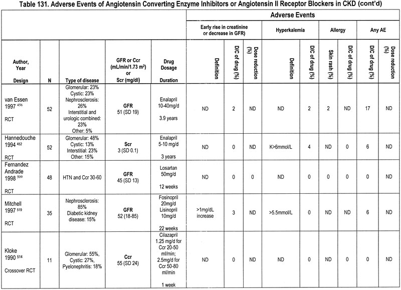
Nkf Kdoqi Guidelines
Mild To Moderate Renal Pelvis Dilatation Identified During

Nomogram Of Fetal Renal Growth Expressed In Length And Parenchymal
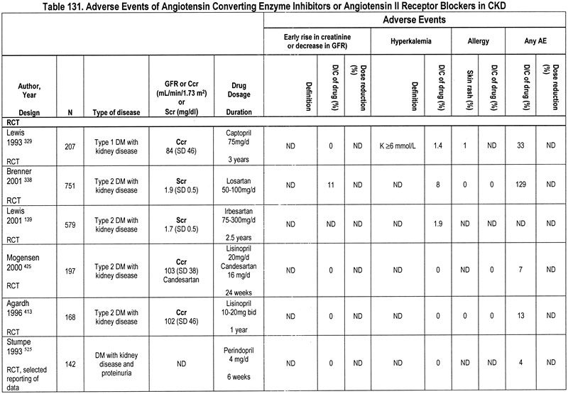
Nkf Kdoqi Guidelines

Age Related Changes Of The Human Fetal Kidney Size Semantic Scholar

Hydronephrosis Ssm Health

The Renal Parenchyma Evaluation Of A Novel Ultrasound Measurement
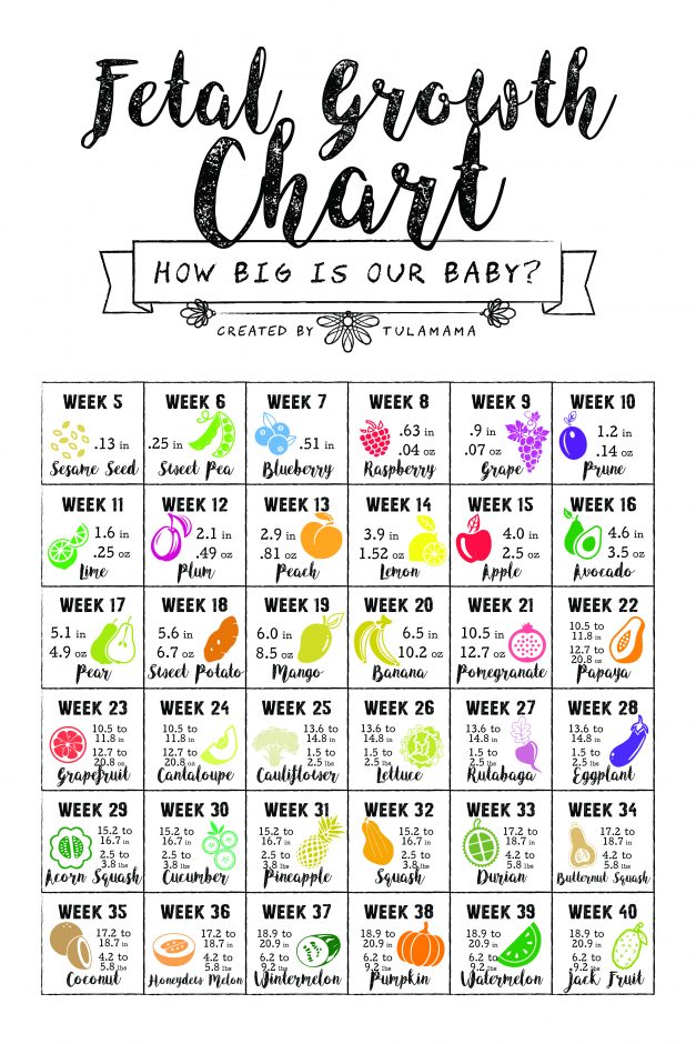
How Big Is My Baby Using A Fetal Growth Chart To Track Your Baby

Charts Of Fetal Size Limb Bones Chitty 2002 Bjog An

Renal Anomalies Detected Or Suspected Antenatally

Predictive Value Of Fetal Renal Artery Doppler Indices In

Conserved And Divergent Features Of Human And Mouse Kidney
Https Www Ajronline Org Doi Pdf 10 2214 Ajr 157 3 1872242
Http Repository Tnmgrmu Ac In 10124 1 220600717karthika Pdf

Evaluation Of Gestational Age By Fetal Occipitofrontal Diameter In
Congenital Solitary Kidney Size At Birth Could Predict Reduced

Maternal Physiologic Renal Pelvis Dilatation In Pregnancy